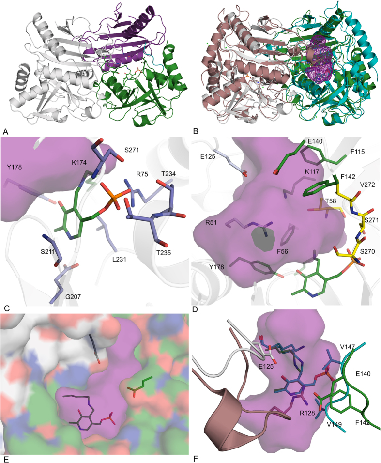
Figure 6. Crystal structure of CpuTA1.
(A) Overview of the CpuTA1 dimer (chain A first domain in magenta, second domain in green, chain B in grey); (B) Overview of a superposition of the CpuTA1 dimer (chain A in green, chain B in grey) with the AT-ωTA dimer (Pdb-code: 4CE5, chain A in turquoise, chain B in brown) with the active site cavity of CpuTA1 depicted as magenta mesh; (C) PLP binding amino acids (blue); (D) active site amino acids (small binding pocket in yellow, large binding pocket in green); (E) entrance tunnel; (F) variable loops of CpuTA1 compared to AT-ωTA. The cavity analysis was calculated by Casox56. The figures were prepared using the program PyMOL (Schrodinger Inc.).

