
Figure 2. Jurkat-Gag cells release biologically active p17.
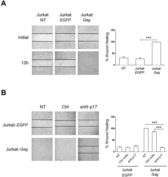
(A) Ability of secreted p17 to stimulate endothelial cell migration. Jurkat, Jurkat-EGFP or Jurkat-Gag cells were added for 16 h to confluent HUVEC monolayers. After several washes with complete medium to remove added cells, HUVEC monolayers were scratched using a 200 μl pipette tip. Cell migration was recorded by light microscopy soon after (initial) and at 12 h after the wound. Pictures are representative of three independent experiments with similar results (magnification 10x). (B) Specificity of p17 biological activity. The same experiment as in (A) was performed in the presence of 1 μg/ml of neutralizing anti-p17 mAb MBS-3 or of an isotype matched control mAb (Ctrl). Neutralizing anti-p17 mAb MBS-3 (anti-p17) −but not an unrelated mAb (Ctrl)−blocked the pro-migratory activity of the p17 secreted by Jurkat-Gag cells. Images are representative of three independent experiments with similar results (magnification 10x). Graphs in the right panels represent quantitative analyses of wound-healing upon different co-culture conditions. Statistical analysis was performed by one-way ANOVA, and the Bonferroni’s post-test was used to compare data (***P < 0.001). NT indicates not treated cells.