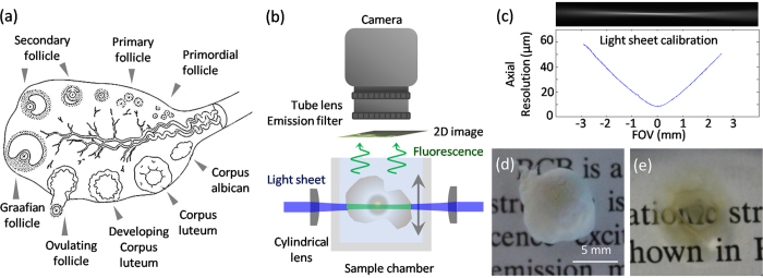
Figure 1. Ovarian follicles and SPIM experimental setup.
(a) Representative sketch of follicle anatomy. Primordial follicles are small, and usually located close to the outer edge of the ovarian cortex surrounded by a single layer of granulosa cells. When the primordial follicle receives hormonal stimulation it forms a primary follicle with two layers of granulosa cells. The primary follicle transitions through the secondary follicle stage, during which small amounts of fluid accumulate in the intracellular spaces. These gradually coalesce to form an antrum, and later the Graafian follicle forms. In uniparous mammals one Graafian follicle ovulates, while the rest degenerate into atretic follicles. Upon pregnancy, the ovulating follicle forms the corpus luteum. The figure was produced, in part, using Servier Medical Art. (b) Side view of the SPIM imaging system. Two light-sheets were spatially overlapped and oriented horizontally. The imaging camera and the imaging optics were placed directly above the sample, which was centrally oriented at the beam waists of the light sheets. By placing the camera orthogonally to the light sheets, 2D fluorescent images through the entire specimen measuring up to 10 mm across could be captured. The samples were placed inside a glass chamber filled with clearing solution and scanned in a linear (vertical) geometry. By translating either the light-sheet or the sample chamber, full 3D image stacks of the sample could be obtained. (c) Light sheet characterisation using a solution of fluorescent dye. The axial resolution across the field-of view (FOV) is determined by the light sheet thickness of 16 μm at the beam waist, expanding to about 60 μm at a distance of 3 mm away from the centre of FOV. (d) Brilliant Cresyl Blue (BCB) stained ovarian follicle. (e) Sample after clearing. Ethanol was used for dehydrating the sample, and 2:1 benzyl alcohol/ benzyl benzoate (BABB) for clearing.

