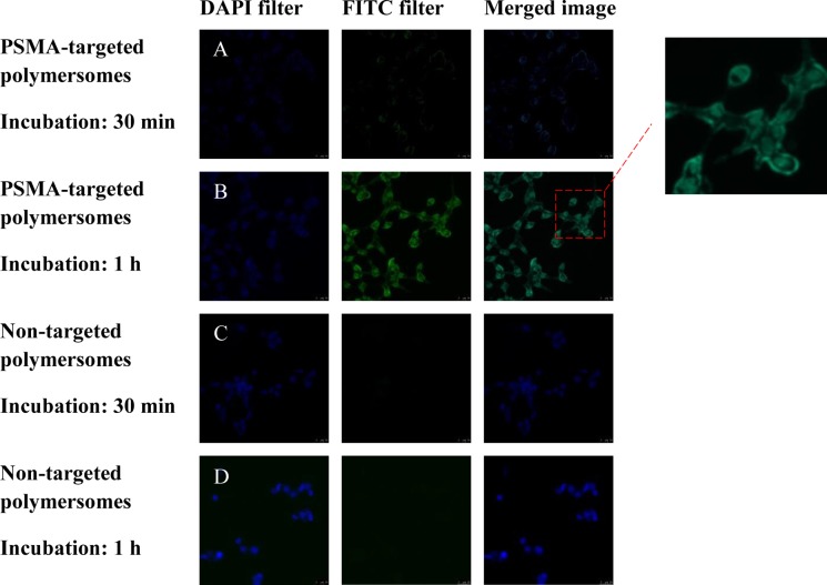
Figure 6.
Fluorescence microscopic images of LNCaP cell incubated with the polymersomes (magnification: 20×). The nuclei of the cells were stained with the Hoechst dye (blue image, 4’,6-diamidino-2-phenylindole filter). The polymersome images are green due to the encapsulated carboxyfluorescein (fluorescein isothiocyanate filter). The merged images are shown in the third panel. (A) PSMA-targeted polymersomes after 30 min incubation, (B) PSMA-targeted polymersomes after 1 h incubation, (C) nontargeted polymersomes after 30 min incubation, and (D) nontargeted polymersomes after 1 h incubation.

