. 2016 Jun 30;7(10):6551–6562. doi: 10.1039/c6sc02588a

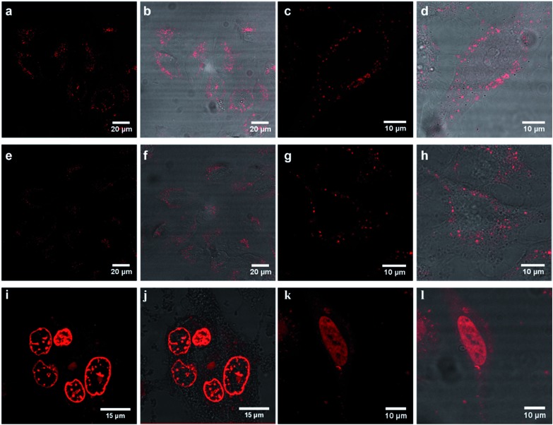
Fig. 3. Confocal luminescence images of (a–d) Ru-ER, (e–h) Ru-Arg8 and (i–l) Ru-NLS in live HeLa cells where the ruthenium channel and overlay channel are shown for each complex. Cells were incubated in the absence of light with 70 μM Ru-ER and Ru-Arg8 for 4 h, and 40 μM with Ru-NLS for 24 h. The distribution of each complex is shown in a group of cells (column 1 and 2) and then focused on a single HeLa cell (column 3 and 4). Ru-ER and Ru-Arg8 were excited using a 488 nm white light laser and the emission was collected between 590 and 700 nm. Ru-NLS was excited using a 470 nm white light laser and the emission was collected between 565 and 700 nm.

Fig. 3