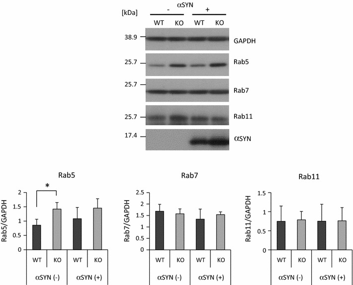
Fig. 5.

Higher Rab5 protein level in KO microglia than in WT microglia. Western analysis was performed to determine the protein levels of Rab5, Rab7 and Rab11 in KO and WT microglia treated with or without αSYN. The quantified density of each Rab was normalized by the density of GAPDH. n = 4 culture wells per group. In all graphical representations, data are expressed as mean ± SD and were assessed by ANOVA (KO vs WT). Rab5: F = 4.28, p = 0.028; Rab7: F = 0.96, p = 0.443; and Rab11: F = 0.01, p = 0.999. The experiment was carried out three times using primary microglia isolated from independent mice, and a representative image and data are shown
