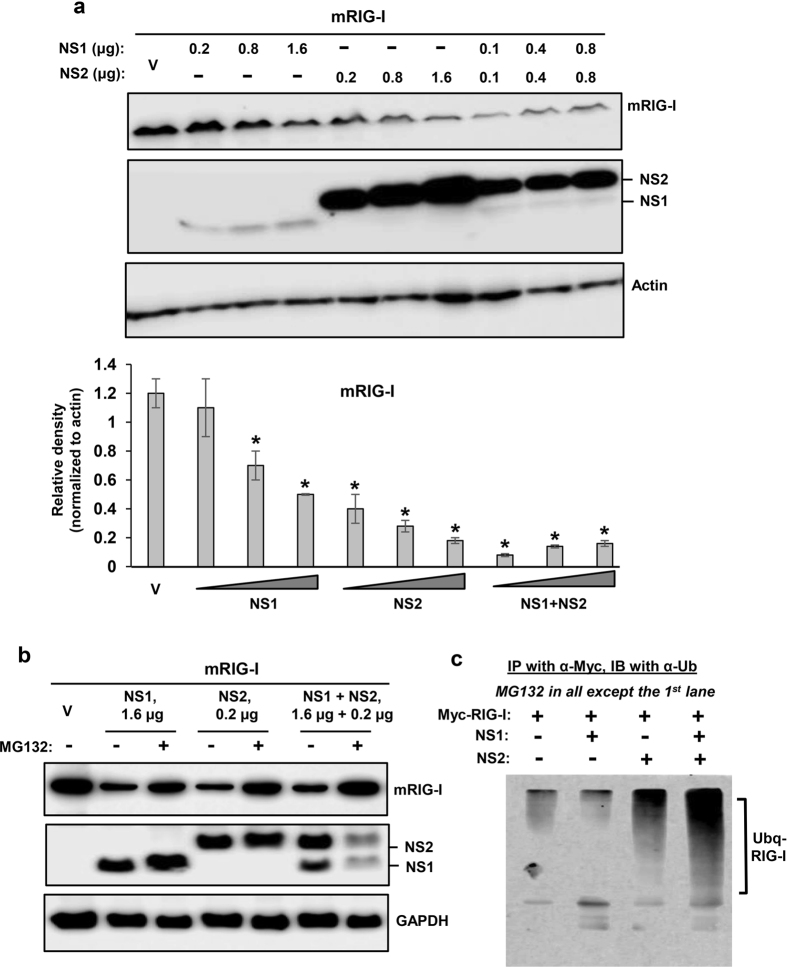
Figure 1. Proteasomal degradation of mouse RIG-I by PVM NS proteins.
MEF cells in 24-well plates were transiently transfected with 0.4 μg Myc-FLAG-mRIG-I expression plasmid and indicated amounts of pCAGGS-FLAG-NS plasmids. Cells were processed 24 h later for immunoblot (Western) to detect the proteins as shown. Myc-FLAG-RIG-I was detected by FLAG antibody. Note that our NS1-expressing plasmid consistently produces less protein than the NS2-expression plasmid, for reasons that are unclear. Cells were cultured in the (a) absence or (b) presence of MG132 (10 μM, added in the medium at 8 h after plasmid transfection), as shown. Actin and GAPDH are loading controls. Densitometry and plot of RIG-I band intensities were performed as described under Methods. V indicates transfection with empty (no-NS) pCAGGS vector. For ubiquitination analysis (c), cells were cultured similarly, immunoprecipitation was performed as described18,19 and the precipitate subjected to immunoblot using pan anti-Ub antibody. The ubiquitinated forms of RIG-I are indicated.

