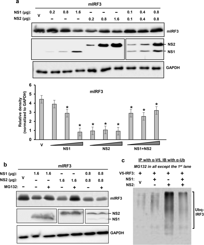
Figure 2. Proteasomal degradation of mouse IRF3 by PVM NS proteins.
These experiments were designed and performed essentially as in Fig. 1, with 0.4 μg V5-mIRF3 replacing the RIG-I expression plasmid. Immunoblot shows the levels of the indicated proteins. As in Fig. 1, cells were cultured in the (a) absence or (b) presence of MG132. GAPDH is loading control. Densitometry and plot of IRF3 band intensities were performed as described under Methods. Ubiquitination analysis (c) was also performed essentially as in Fig. 1c, and the ubiquitinated forms of IRF3 are indicated.

