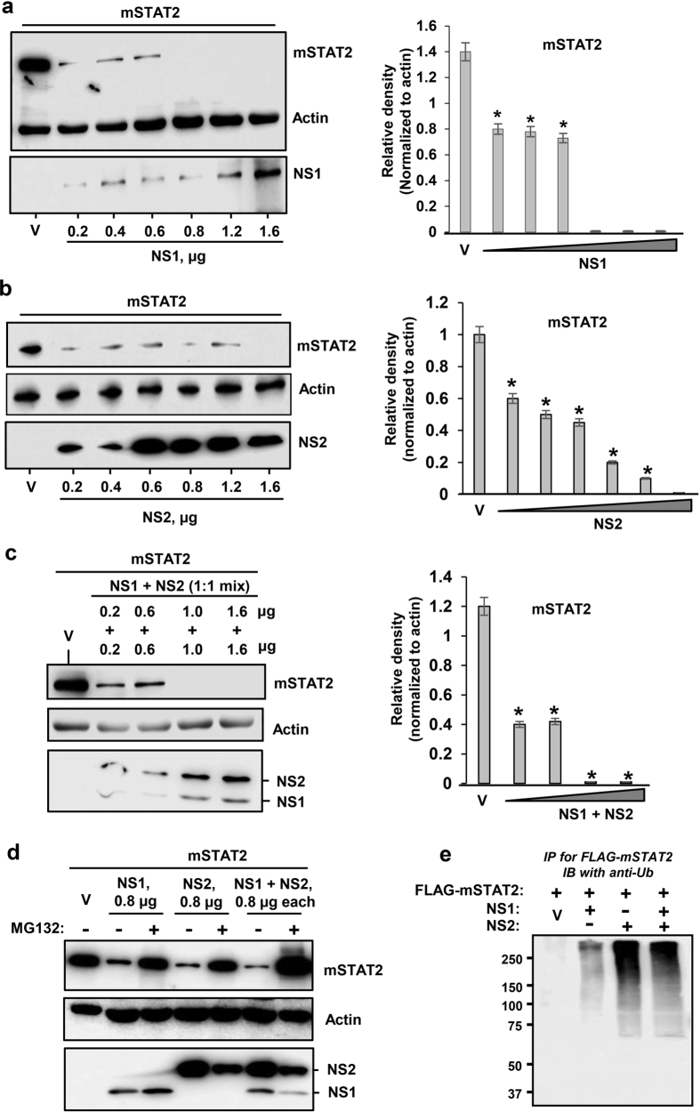
Figure 4. Degradation of mouse STAT2 by PVM NS proteins.
Transient transfection was performed as in Fig. 1 to express FLAG-mSTAT2 along with (a) NS1 protein, (b) NS2 protein or (c) both. Right panels show densitometric plot of the corresponding substrate bands on the left. Strong reduction of STAT2 is observed in all three panels (a,b,c). Actin is loading control. V indicates 0.4 μg vector (pCAGGS, no NS). (d,e) PVM NS proteins promote ubiquitination and proteasomal degradation of mSTAT2. These experiments are essentially similar to Fig. 1c, FLAG-mSTAT2 being the substrate here. Protection of STAT2 by MG132 (d) and the ubiquitinated form of STAT2 (e) are demonstrated. Where shown, ‘V’ indicates vector-only (no NS), and actin is the loading control.

