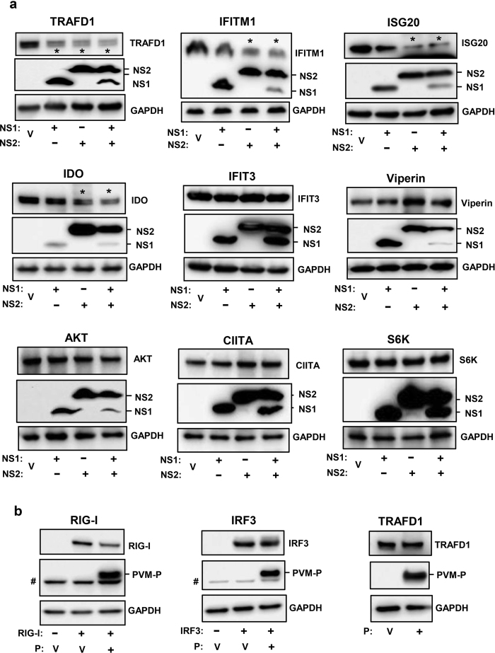
Figure 7. Degradation of specific ISGs by PVM NS proteins but not by PVM P protein.
(a) Tet-induced FLAG-ISG-expressing cells were transfected as described in Methods with 1.6 μg FLAG-NS1 or 0.2 μg FLAG-NS2 plasmid or both, and cells were harvested for immunoblot analysis using FLAG antibody as primary antibody to detect the ISG and NS proteins. Four ISGs (TRAFD1, IFITM1, ISG20, IDO), affected by NS proteins (of various combinations), are shown first, followed by two ISGs (IFIT3, Viperin) that are representative of many that were not affected. Three proteins, not related to IFN pathways (AKT, CIITA, S6K), were also not affected. (b) MEF cells were transfected with 1.6 μg of FLAG-RIG-I or V5-IRF3 plasmids (as in Figs 1 and 2, respectively), along with FLAG-PVM-P (1.6 μg) plasmid where indicated. For TRAFD1, the Tet-inducible clone was induced with Tet (as in panel ‘a’). A nonspecific band of MEF origin, migrating just below the P band, is marked by #; it is not seen in the HEK293 cells. In all panels, V indicates transfection with empty pCAGGS vector (i.e. no NS or P). The IFITM1 gel was 12% polyacrylamide; all others were 4–20% gradient.

