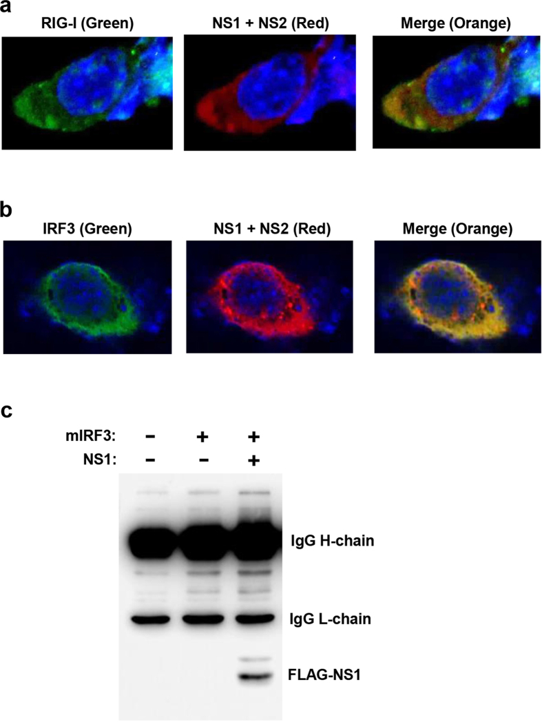
Figure 8. Interaction of NS proteins with their cellular substrates.
(a,b) Immunofluorescence microscopy, performed as described in Methods, show co-localization of PVM NS proteins (Red) with substrates RIG-I or IRF3 (Green), indicated by orange colours in the merged panel. Nuclei are DAPI-stained (Blue). (c) Immunoprecipitation (IP) of V5-IRF3 using mouse anti-V5 antibody, followed by immunoblotting with mouse anti-FLAG antibody (which provides cleaner background than the corresponding rabbit antibody), detected FLAG-NS1 as shown. The dark upper bands are heavy and light chains of the immunoprecipitating V5 antibody, detected by the secondary antibody. In both microscopy and IP, MG132 was added to the cell culture to stabilize the substrates, so that the substrate-NS complexes could be detected.

