Figure 1. Kir2.1 at the ID.

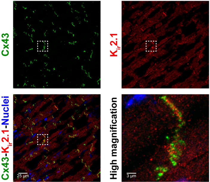
Representative confocal images of A) Cx43, B) Kir2.1 and C) overlay demonstrate co-localization of Cx43 and Kir2.1 in ventricular sections. D) A close up view demonstrates enrichment of Cx43 and Kir2.1 at the ID.

Representative confocal images of A) Cx43, B) Kir2.1 and C) overlay demonstrate co-localization of Cx43 and Kir2.1 in ventricular sections. D) A close up view demonstrates enrichment of Cx43 and Kir2.1 at the ID.