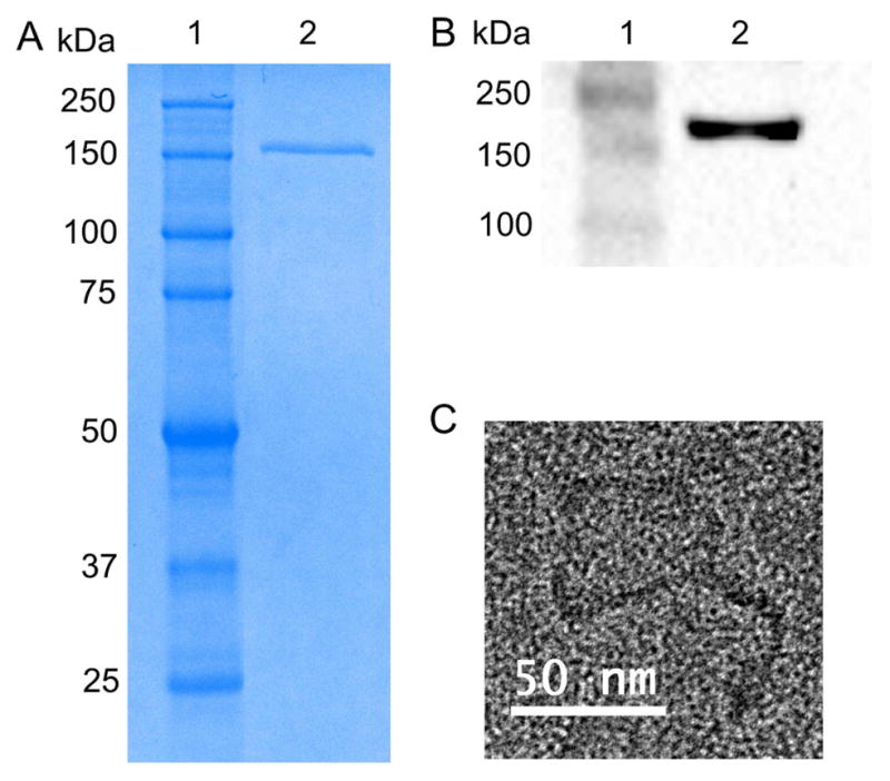
Figure 1.

Clathrin isolation from bovine brains. (A) SDS-PAGE of the triskelion sample (MW ~180 kDa) on a 9% gel: ladder (lane 1), clathrin triskelion (lane 2). (B) A western blot analysis confirms the presence of clathrin in the sample. An anti-clathrin heavy chain antibody was used to detect the clathrin triskelion: ladder (lane 1), clathrin triskelion (lane 2). (C) TEM image of a clathrin triskelion.
