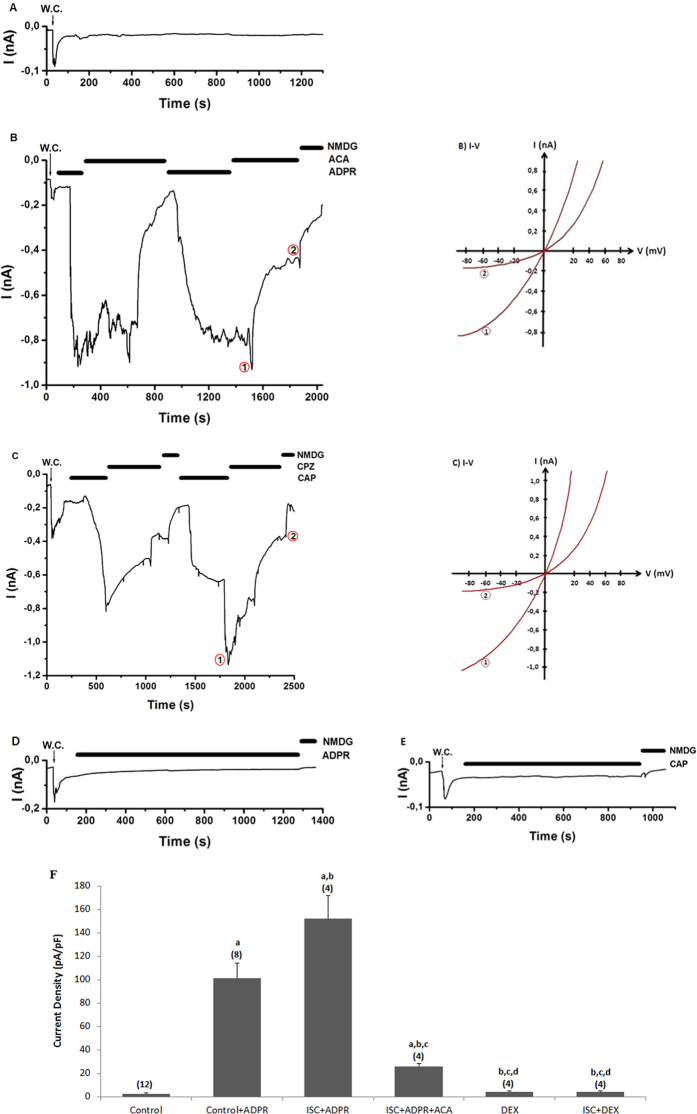
Figure 3. Effects of dexmedetomidine (DEX) treatment on TRPM2 channel activation in the DRG of in control and cerebral ischemia (ISC)-induced rats.
The holding potential was minus sixty millivolt in the analyses. (A) Control: Original recordings from control neuron. (B) Control+ADPR group: DRG isolated from rats of control and sham groups without ISC induction and they were stimulated by capsaicin (0.01 mM) and ADPR (1 mM) but inhibited by ACA (0.025 mM). (C) ISC+ADPR group: TRPM2 currents in the DRG neurons of ISC-induced rats were gated by ADPR (1 mM) in the patch pipette and they were inhibited by ACA in the bath of patch chamber. (D) DEX+ADPR group: The rats received intraperitoneal DEX and then the DRG neurons were stimulated by in vitro ADPR (1 mM in patch pipette). (E) ISC+DEX+ADPR group: The rats received DEX at 3rd, 24th and 48th hours after induction of experimental ISC and then the DRG was stimulated by ADPR (1 mM). (B) I-V and (C) I-V: Current voltage relationships and they are same experiments as in panels B and C, respectively. W.C. is whole-cell. (F) TRPM2 channel capacitance of the DRG in control and ISC-induced rats (mean ± SD). The DRG neurons were further treated in vitro afterwards with ADPR (1 mM) and ACA (0.025 mM). Capacitance calculation of the currents was described in the method section. The numbers of group were indicated in parentheses. (ap ≤ 0.001 versus control. bp ≤ 0.001 versus control+ADPR group. cp ≤ 0.001 versus control+ADPR+ACA group. cp ≤ 0.001 versus ISC+ADPR group. ep ≤ 0.001 versus ISC+ADPR+ACA group).

