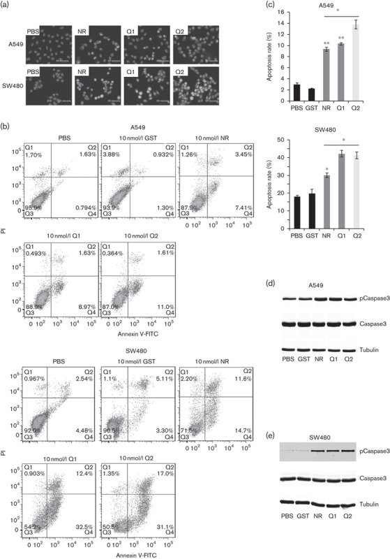
Fig. 5.

Effects of RGD-IFN-α2a-core fusion proteins on tumor cell apoptosis induction. (a) Hoechst 33342 staining. Cells were seeded onto coverslips and treated with 10 nmol/l of NR, Q1, or Q2 for 24 h and then subjected to Hoechst 33342 staining. (a–d) A549 cells treated with PBS, NR, Q1, or Q2; (d, e) SW480 cells treated with PBS, NR, Q1, or Q2. (b) Flow cytometric assay. The cells described above were subjected to flow cytometric assay after the treatments described above. (c) Summarized data of B. The data are expressed as means±SEM from at least three independent experiments. *P<0.05 and **P<0.01 compared with the control. (d) Western blot analysis of caspase-3 levels in A549 cell lines. Duplicated cells were subjected to western blot analysis. The values of PBS-treated, GST-treated, IFN-α2a-treated, Q1-treated, and Q2-treated tumor cells were expressed as means±SEM (n=5). (e) Western blot analysis of caspase-3 levels in SW480 cell lines. Duplicated cells were subjected to western blot analysis. The values of PBS-treated, GST-treated, NR-treated, Q1-treated, and Q2-treated tumor cells were expressed as means±SEM (n=5). GST, glutathionine S-transferase; IFN, interferon; NR, nitrate reductase; PI, propidium iodide; RGD, arginine–glycine–aspartic acid.
