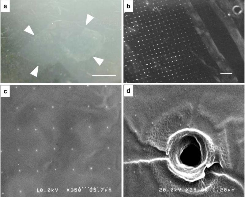
Fig. 3. Characterization of PLLA film.
a) Film floating on water surface after the PVA template dissolved (film edges highlighted with white arrows, scale bar 1 cm); b) optical microscope image of the film (holes appear as white dots, scale bar 500 μm); c) the array of holes in the film are visualized with the SEM (2 nm gold coating); d) A detail of a single hole in the PLLA film.

