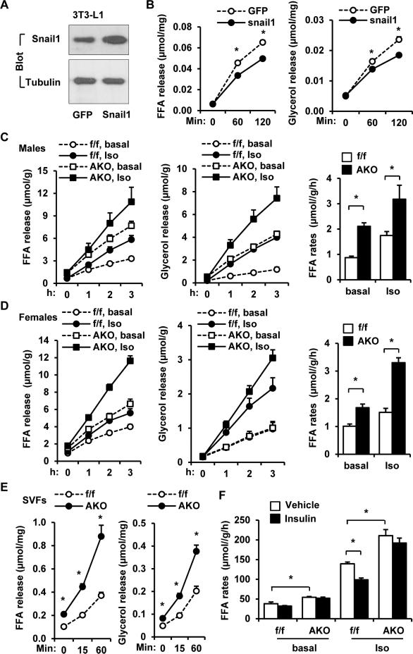
Figure 2. Snail1 suppresses adipocyte lipolysis.
A-B. 3T3-L1 adipocytes were infected with Snail1 or GFP adenoviruses for 2 days. A. Cell extracts were immunoblotted with antibodies against Snail1 or tubulin. B. Adipocytes were stimulated with isoproterenol (1 μM), and FFA and glycerol releases were measured (normalized to protein levels). n=3. C-D. Gonadal WAT explants were prepared from male (12 weeks) (AKO: n=3; Snail1flox/flox: n=5) and female (20 weeks, n=4) mice, and stimulated with or without isoproterenol (1 μM). FFA and glycerol release rates were measured (normalized to protein levels). FFA release rates (curve slopes) were calculated. E. SVFs were prepared eWAT, differentiated into adipocytes, and stimulated with isoproterenol (1 μM). FFA and glycerol release rates were measured. n=6. F. EMSCs were isolated from Snail1flox/flox and AKO males, differentiated to adipocytes, and stimulated with PBS vehicle or insulin (100 nM for 6 h) in the absence (basal) or presence of isoproterenol (0.1 μM). FFA release rates were measured (normalized to protein levels). The values are mean ± sem. *p<0.05.

