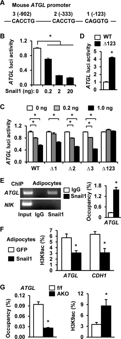
Figure 4. Snail1 epigenetically represses the ATGL promoter.
A. A schematic representation of the mouse ATGL promoter (numbers: positions from the transcription start site). B. ATGL luciferase reporter plasmids were cotransfected with Snail1 expression vectors into HEK293 cells. Luciferase activities were measured 48 h after transfection and normalized to β-gal internal control. C. WT or mutant ATGL luciferase reporter plasmids were cotransfected with Snail1 plasmids into HEK293 cells, and luciferase activities were measured 48 h after transfection. D. WT or Δ123 ATGL luciferase reporter plasmids were transfected into 3T3-L preadipocytes. Luciferase activities were measured 48 h after transfection and normalized to β-gal internal control. E. ChIP assays were performed using 3T3-L1 adipocytes. Snail1 occupancy on the ATGL promoter (normalized to inputs) was quantified by qPCR. F. 3T3-L1 adipocytes were infected with Snail1 or GFP adenoviruses for 24 h. ATGL and CDH1 promoter H3K9 acetylation levels (normalized to inputs) were quantified by ChIP-qPCR. G. Gonadal WAT was isolated from Snail1flox/flox (n=3) and AKO (n=3) females fed a HFD for 8 weeks. Snail1 occupancy and H3K9 acetylation levels in the ATGL promoter were measured by ChIP-qPCR. The values are mean ± sem. *p<0.05.

