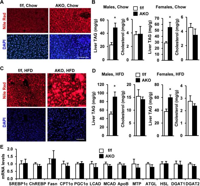
Figure 7. Adipocyte-specific deletion of Snail1 promotes hepatic steatosis.
A-B. Mice (8 weeks) were fed a normal chow diet. A. Nile red staining of male liver sections (overnight fasting). Scale bar: 100 μm. B. Liver TAG and cholesterol levels (normalized to liver weight). AKO males: n=3, Snail1flox/flox males: n=5, AKO females: n=7, Snail1flox/flox females: n=7. C-D. Mice were fed a HFD for 8 weeks (females) or 30 weeks (males). C. Nile red staining of male liver sections (overnight fasting). Scale bar: 100 μm. D. Liver TAG and cholesterol levels (normalized to liver weight). AKO (n=17) and Snail1flox/flox (n=19) males were fasted for 24 h; AKO (n=3) and Snail1flox/flox (n=6) females were randomly fed. E. Gene expression (normalized to 36B4 expression) was measured in the liver by qPCR. The values are mean ± sem. *p<0.05.

