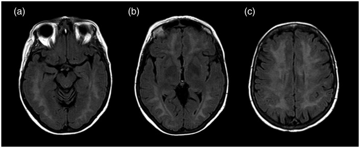
Figure 1.
T2 FLAIR (FLuid Attenuated Inversion Recovery) on axial plan at three different levels: (a) hyppocampal cortex; (b) globus pallidus; (c) semioval center at 3 days from CO poisoning. The figure shows a widespread increase in T2 signal intensity at bi-hemisferic periventricular white matter without involvement of the basal ganglia structures. However, this alteration of white matter intensity is not specific to CO poisoning.

