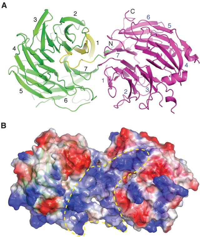
Figure 1.

Overall structure of the WD40 repeat domains of Gemin5. (A) Ribbon diagram showing that an N-terminal short β strand completes the C-terminal β-propeller structure (magenta) and showing four following β strands (yellow) form the first blade of the N-terminal seven-bladed β-propeller structure (green). (B) The surface electrostatic potential of G5N shows a continuous positively charged region (enclosed in the yellow dashed line) potentially capable of binding RNA. The partially transparent surface is superimposed with the ribbon representation of G5N and is viewed from a direction similar to that in A.
