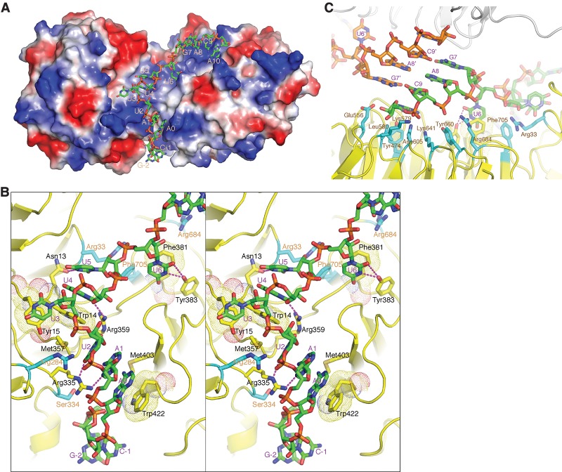
Figure 3.
G5N–RNA cocrystal structure. (A) The 13-mer U4 snRNA fragment is bound in the positively charged region at the interface of the tandem WD40 repeat domains and the C-terminal WD40 domain. The G5N structure is shown in a surface representation colored according to its surface electrostatic potential ([blue] positive; [red] negative; [white] neutral) and is viewed from the same direction as in Figure 1A. The RNA is shown in a stick model ([green] carbon; [blue] nitrogen; [red] oxygen; [orange] phosphorus) and is numbered as in Figure 2A. (B) A stereo diagram showing the interaction of the first 9 nt of RNA with G5N. The involved amino acid residues are shown in a stick model, with the carbon bonds involved in contacting RNA bases colored yellow, and the carbon bonds (labeled with orange letters) involved in non-base-contacting interactions colored cyan. Hydrogen bonds are indicated with magenta dashed lines, and aromatic residues involved in stacking with RNA bases are superimposed with a dot representation. (C) G5N–RNA interactions involving the last 4 nt. A neighboring G5N–RNA within the same asymmetric unit is shown with the protein in a gray ribbon representation, and the RNA is shown in a stick model with the bonds connected to phosphorus atoms colored orange.

