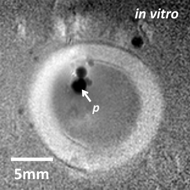
Figure 3a:

(a) Transaxial MR image of a porcine aorta acquired in vitro from the probe (p) (three-dimensional turbo spin-echo sequence, 1500/16 [repetition time msec/echo time msec]; voxel-size, 0.15 × 0.15 × 2 mm; duration, 170 seconds). (b) Temperature map produced with MR thermometry (two-dimensional gradient-echo sequence, 50/25; flip angle, 25°; voxel size, 0.25 × 0.25 × 6 mm; duration, 8 seconds) relative to a 25°C baseline temperature. The thermally affected region is annotated (dashed area). The artifact in the top left (dashed arrow) above the heated area is attributed to increasing thermal convection away from the ablation site, causing signal dephasing over the time frame of this image acquisition. (c) Temperature map acquired with MR thermometry (two-dimensional gradient-echo sequence, 41/25; flip angle, 24°; voxel size, 0.3 × 0.3 × 8 mm; and duration, 6 seconds) in the target area from Figure E4a (online), overlaid on the MR image.
