
Figure 3.
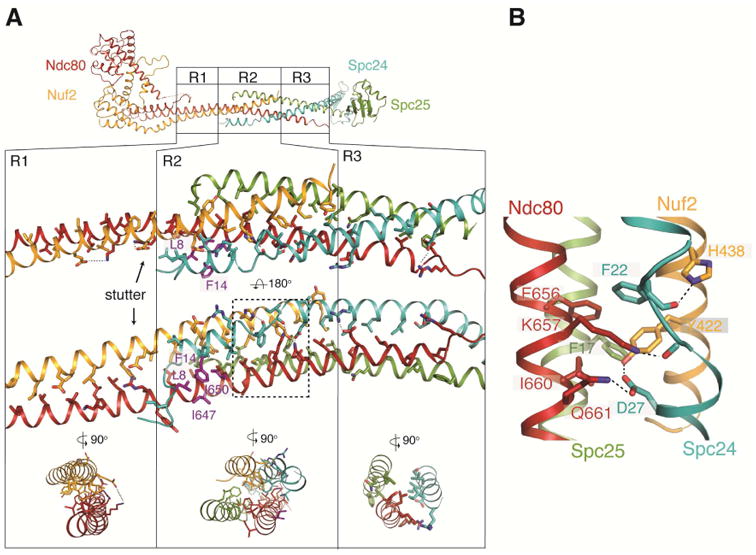
Central region of Ndc80cdwarf/e-dwarf. (A) Top: Diagram of Ndc80ce-dwarf; regions R1, R2 and R3 are identical in the Ndc80cdwarf and Ndc80e-dwarf structures (Cα rmsd = 0.8 Å). Middle: Expanded view of R1, R2 and R3. Arrow points to an additional, “stutter” residue in the third heptad in R1. Residues shown as purple sticks (Ndc80: I650 and I647; Spc24: L8 and F14) form a hydrophobic surface that contacts the nanobody (Figure S3). Bottom: cross section diagrams to show two-chain, four-chain, and three-chain packing in R1, R2 and R3, respectively. (B) Expanded view of a network of aromatic and ionic interactions, conserved in fungi. See also Figure S3.