
Figure 4.
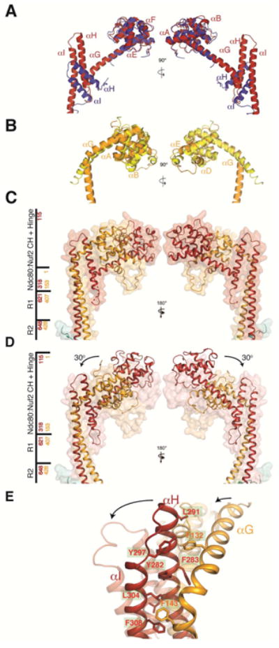
Comparison of yeast and human Ndc80 and Nuf2 CH domains and Ndc80c hinge. (A) Superposition of the Ndc80 CH domain from human (in blue; coordinates from chain B of PDB entry 2VE7) and yeast (in red, chain A of the Ndc80cdwarf crystal structure). (B) Superposition of Nuf2 CH domains from human (in yellow; chain D of PDB entry 2VE7) and yeast CH domain (in orange; chain B of the Ndc80cdwarf crystal structure). (C) Structure of CH domains and adjacent coiled coil from Ndc80ce-dwarf as both secondary-structure ribbons and transparent surface. Side bar marks regions of the structure; red and yellow numbers correspond to Ndc80 and Nuf2, respectively. (D) Structure of Ndc80cdwarf juxtaposed on transparent surface of Ndc80ce-dwarf. The arrows show the reorientation of the Ndc80:Nuf2 CH domains with respect to the coiled-coil axis. Structures aligned with respect to residues encompassing the R1 and R2 segments of the middle region with an RMSD of 0.8 Å2. (E) Fulcrum of hinge, showing cluster of aromatic residues. See also Figure S3E and Supplemental Movie 1.