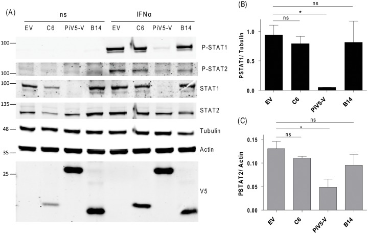
Fig 2. C6 does not inhibit the IFNα-induced phosphorylation of STAT1 or STAT2.
HeLa cells stably expressing the proteins shown were stimulated with IFNα (1000 U/ml for 1 h). Cells were harvested and cell lysates subjected to SDS-PAGE and immunoblotting to assess levels of STAT1 and STAT2 phosphorylation (A). Samples were also immunoblotted for alpha tubulin and actin as controls. Positions of molecular mass markers are shown on the left of the figure. Quantification of triplicate samples of phosphorylated STAT1 (B) and phosphorylated STAT2 (C) proteins was performed using Odyssey software (LICOR) and are shown relative to a constant house-keeping gene. P<0.05. Immunoblots were performed at least twice and a representative figure is shown.

