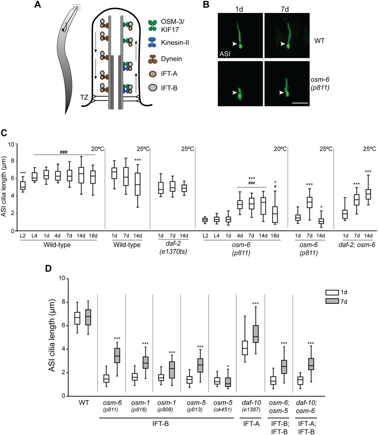
Fig 1. Cilia of the ASI sensory neurons elongate in aged IFT mutants.
(A) (Left) Cartoon of a worm showing a representative sensory neuron in the worm head. Cilia are present at the dendritic ends at the nose (box). (Right) Diagrammatic representation of the structure of a typical cilium and IFT in C. elegans. Arrows indicated direction of IFT. TZ—transition zone (showing Y-link microtubule-to-membrane connectors). (B) Representative images of ASI cilia in 1d and 7d old adult wild-type (WT) and osm-6(p811) mutants. Arrowheads indicate the cilia base. Anterior is at top. ASI cilia were visualized via expression of a GFP-tagged SRG-36 GPCR protein expressed under the ASI-specific str-3 promoter. Scale bar: 5 μm. (C) Quantification of ASI cilia length in animals of the indicated genetic backgrounds at different larval stages (L2, L4) or days of adulthood. Horizontal lines indicate 25th, 50th and 75th percentiles; bars indicate 5th and 95th percentiles. * and *** indicate different from 1d within a genotype at P<0.05 and 0.001, respectively; # and ### indicate different from L2 within a genotype at P<0.05 and 0.001, respectively (Kruskal-Wallis test with post hoc paired comparisons). n>30 for each; ≥3 independent experiments. Animals were grown at either 20°C or 25°C for each set of experiments (indicated at top right). (D) Quantification of ASI cilia length in 1d and 7d old animals of the indicated genotypes grown at 20°C. ASI cilia were visualized via expression of str-3p::srg-36::gfp. Alleles used in the double mutant strains were osm-6(p811), osm-5(p813), and daf-10(e1387). Horizontal lines indicate 25th, 50th and 75th percentiles; bars indicate 5th and 95th percentiles. * and *** indicate different from 1d within a genotype at P<0.05 and 0.001, respectively (Wilcoxon Mann-Whitney U test). n>30 for each; ≥3 independent experiments.

