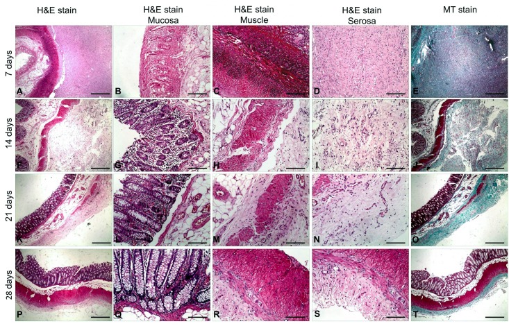
Fig 5. Histopathology of the colon wall after irreversible electroporation (IRE) ablation.
(A-E) The colon wall shows necrosis in the mucosa, submucosa and muscle layers 7 days after IRE ablation and significant fiber proliferation in the serosa layer (E). Inflammatory cell infiltration (C) and significant proliferation of serosa layer fibers (D-E) can also be observed. (F-J) Fourteen days after IRE ablation, the colon wall shows hyperplasia of the mucosal glands (G), degeneration and necrosis of the muscular layer (H), thickening and fibrosis of the serosa (I, J). (K-O) Twenty-one days after IRE ablation, the muscle layer was almost absent (K), replaced with fibrous granulation tissue (N). (P-T) Twenty-eight days after IRE ablation, the colon wall appears histopathologically normal, with the muscle layer regenerated (R) and the serosa returned to normal thickness (S, T). Scale bars in A, E, F, J, K, O, P and T = 2.5 mm (20× magnification). Scale bars in B–D, G–I, L–N and Q–S = 500 μm (100× magnification). Hematoxylin and eosin stain (H&E stain), Masson’s trichrome stain (MT stain).

