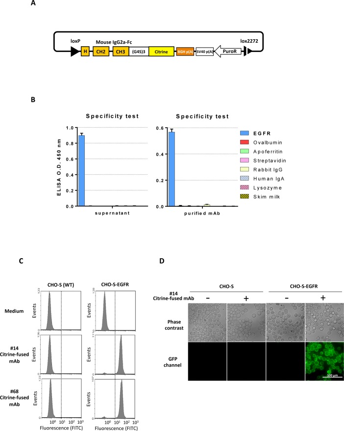
Fig 6. Generation of antibodies fused with a fluorescent protein.
A) Design of pAbCitP donor vector to generate a Citrine-fused mouse chimeric mAb by RMCE. B) ELISA analysis to confirm the specificity after fusion of fluorescent protein. The culture supernatant (left) and purified antibody (right) were used. Negative control antigens are identical to those described in Fig 4B. C) Flow cytometry analysis of CHO-S cells. CHO-S cells overexpressing EGFR (right), and wild-type parental CHO-S cells (left), were incubated in the culture supernatant of the Citrine-fused anti-EGFR mAb expressing cells. Culture supernatants from two independent clones, #14 (middle) and #68 (lower) were examined. Culture medium of parental CHO-S was used as a negative control (upper). D) Fluorescence microscopy analysis using Citrine-fused mAbs. CHO-S cells overexpressing EGFR (right four panels) and parental CHO-S cells (four leftmost panels) were incubated in the culture supernatant of Citrine-fused anti-EGFR mAb-expressing cells and analyzed by fluorescence microscopy. The upper panels are phase contrast images and the lower panels are the images of GFP channel.

