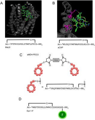
Figure 4.

Examples of macrocyclic‐peptide ligands identified using the RaPID system. (A) Crystal structure of MaL6:PfMATE (PDB: 3WBN) and the sequence of MaL6. MaL6 is represented in stick format and PfMATE is represented in cartoon format. (B) Crystal structure of aCAP:CmABCB1 (PDB: 3WMG) and the sequence of aCAP. aCAP is represented in stick format and a single monomer unit of CmABCB1 is represented in cartoon format. CmABCB1 residues involved in specific interactions with aCAP are coloured magenta. Hydrogen bonds are shown in yellow dashes. (C) Schematic representation of a Met‐binding dimer‐macrocylic‐peptide, aMD4‐PEG3. Figure adapted from Ref. 10. (D) EpCAM‐binding fluorescent macrocyclic‐peptide Epi‐1‐F. X‐ray crystal structures were rendered in PyMOL v1.5.0.4
