
Figure 1.
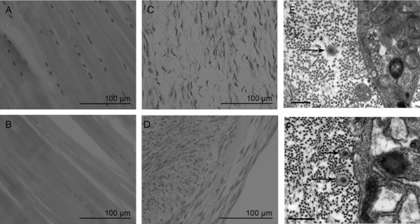
Ultrastructural images of native T/L and 3D TE constructs. H&E staining of native ligament (A), native tendon (B), 3D ligament constructs (C), and 3D tendon construct (D) (Bar 100 μm). Transmission electron microscopy of 3D TE tendon (E) and ligament (F) constructs indicate the presence of aligned extracellular collagen fibrils and fibripositors (black arrows) demonstrating that constructs have formed correctly.