
Figure 2.
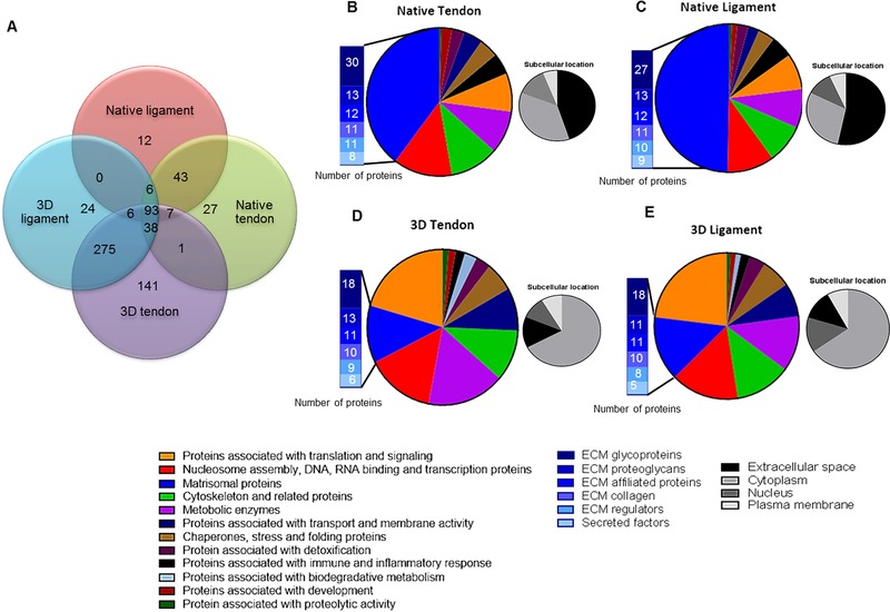
Protein composition of native T/Ls and 3D TE construct identified with PEAKS. The total number of proteins identified following MS in each tissue type as well as common proteins between the tissue types is presented (A). The proteomes of native tendon (B), ligament (C), 3D tendon (D), and 3D ligament (E) constructs were subdivided based on Uniprot function and matrisomal classification (Matrisome Project). The associated subcellular locations of the proteins are also shown (B–E).