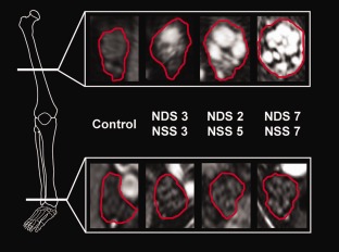
Figure 1.

Array of representative source images indicating segmented tibial fascicles within the sciatic nerve at the thigh level (upper row) and their continuation as the tibial nerve distally at the ankle level (lower row). Nerve lesions are conspicuous under hyperintense signal alteration, which was found to be an effect of increased proton spin density by quantitative signal analysis. Within symptomatic diabetic polyneuropathy (DPN) groups (mild/moderate or severe), there is a strong proximal focus of lesion predominance located at the thigh level (the white reference line in the bony anatomical scheme on the left indicates the slice position). A strong proximal‐to‐distal lesion gradient with a marked decrease at the distal ankle level (lower white reference line at the ankle level) and increasing fascicular involvement across groups of increasing DPN severity (from left to right) were observed. NDS = Neuropathy Disability Score; NSS = Neuropathy Symptom Score.
