
Figure 3.
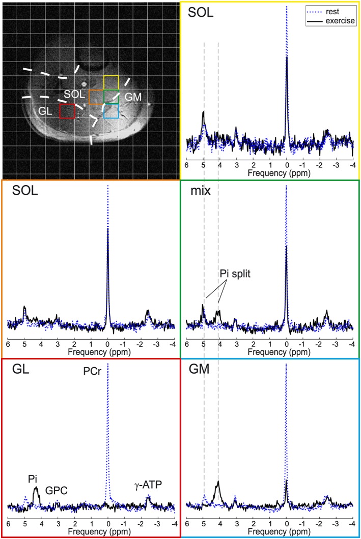
Representative 31P MR spectra acquired at rest (blue dotted line) and at the end of exercise (black solid line) in voxels representing single muscles, i.e. GL, GM and SOL, or a mixture of GM and SOL tissue. Note the Pi splitting in the mixed (green) voxel. A matched filter (6 Hz Lorentzian) was applied for visualization purposes only