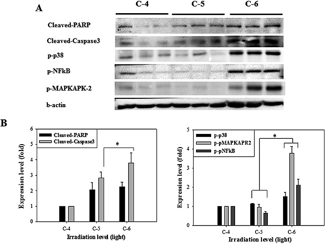
Figure 2.

(A) The result of Western blot analysis on infarct lesion induced by C‐4, C‐5 and C‐6 irradiation. On 30 days, each mouse brain was lysed and detected using apoptosis and inflammatory response‐related proteins. The proteins were significantly phosphorylated in C‐6 irradiation group. (B) Quantitative analysis of expression level in comparison to β‐actin, an internal control. Each bar represents the mean ± SD of independent experiment performed in triplicate (n = 3; *P < 0.05).
