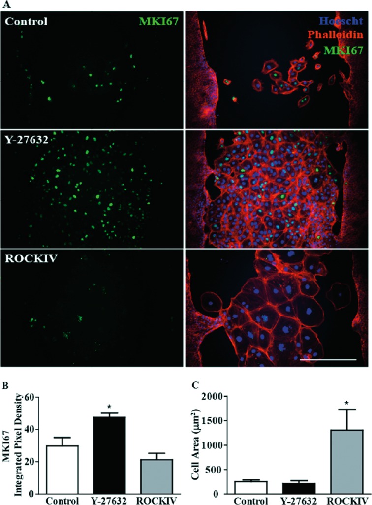
Figure 5.
PanROCK inhibition promotes proliferation in wounded area. (A) Five days after scratch, cells were stained with anti-MKI67 (marker of proliferation KI67, green), phalloidin (F-actin, red), and Hoescht (DNA, blue). Scale bar, 100 μM. (B) MKI67 fluorescence in wounded area was significantly higher when cells were treated with 10 μM of Y-27632. *P ≤ 0.05 to control (C) ROCKIV treatment increased cell size compared with control and Y-27632 treatment. *P ≤ 0.05 to control. Error bars represent ± SEM (n = 3).

