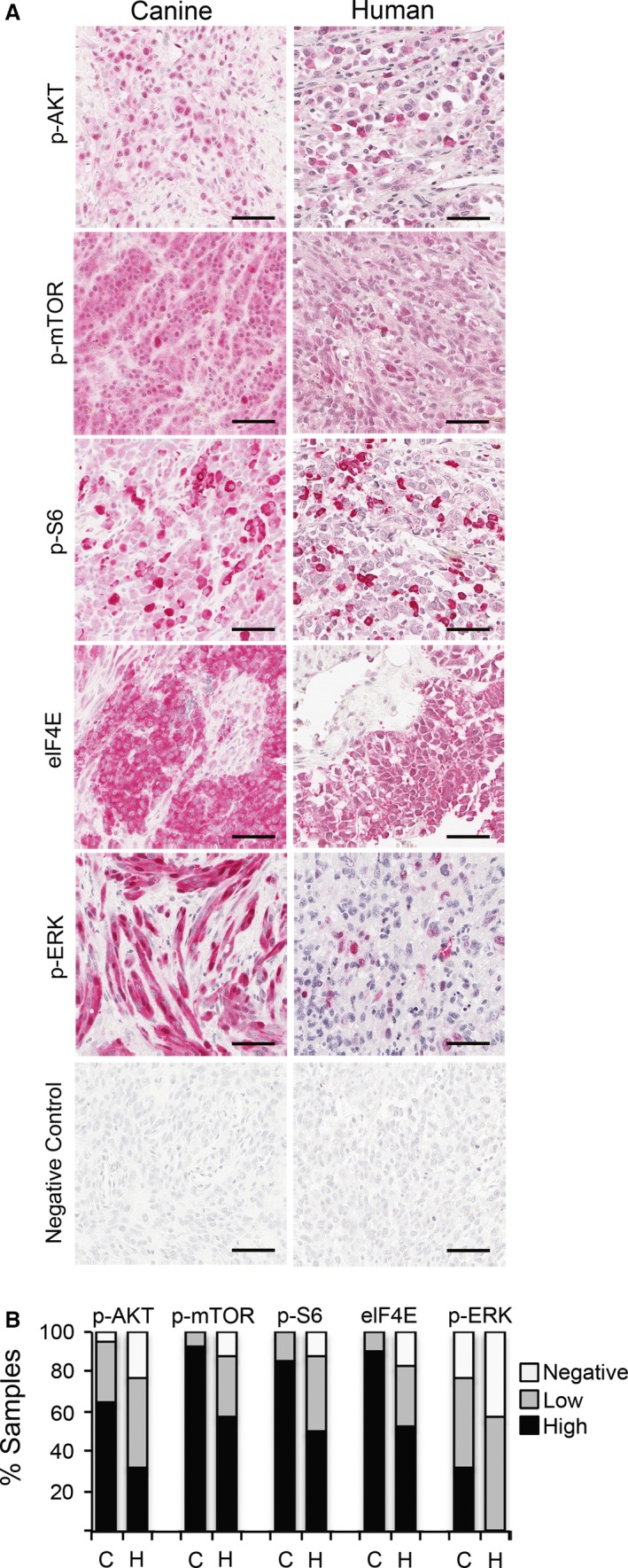
Figure 1.

RAS/ERK and PI3K/AKT/mTOR signal transduction pathway activation in human and canine mucosal melanoma detected by immunohistochemistry on tissue arrays. (A) Representative immunopositive reactions (red chromogen, hematoxylin counterstain) for multiple pathway mediators are illustrated, as labeled. Bars = 50 μm. (B) Percentages of immunopositive melanomas for selected canine (C, n = 43) and human (H, n = 40) signal transduction mediators. Relative intensity and percentages of immunolabeled cells were considered in scores, assigned as negative, low, and high (see Methods).
