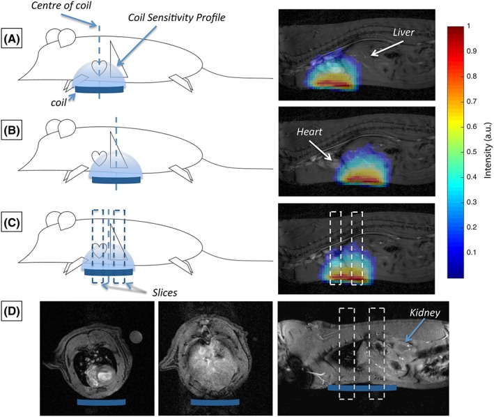
Figure 1.

Illustrative representation of the RF coil placement (and associated coil sensitivity profiles) for the three different acquisition schemes. (A), A global acquisition is used and localization of signal from the heart is achieved solely by the placement of the coil under the heart. (B), A global acquisition is used and localization of signal from the liver is achieved solely by the placement of the RF coil under the liver. (C), Data are sequentially acquired from the heart and liver through the use of a slice‐selective protocol and placement of the RF coil between the heart and the liver. (D), Example axial and sagittal profiles through the rat heart and liver at the levels of the slices acquired in the spectroscopy data obtained with Protocol C, which indicate minimal contamination or contribution from other organs (e.g. the kidneys) at these positions
