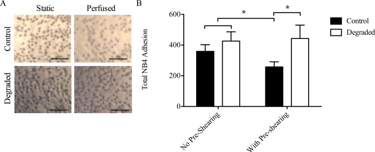
Fig 4. NB4 cell adhesion in 3-D cell culture models with degradation and flow.
(A) Representative light microscope images of NB4 cells adhered to HAEECs in models. Images were acquired at 10x magnification (scale bars = 25μm). (B) Quantification of adherent NB4 cells (n = 3, *p<0.05, error bars represent SEM).

