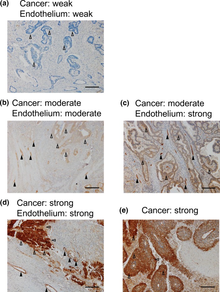
Figure 1.

Representative immunohistochemical staining of JAG1 expression in human colorectal cancer tissues (original magnification ×100, scale bars represent 0.25 mm). (a) Example of cancer and endothelium tissue with weak intensity of staining (jcIHC‐W, jeIHC‐W). (b) Example of cancer and endothelium with moderate intensity of staining (jcIHC‐M, jeIHC‐M). (c) Example of cancer and endothelium with moderate and strong intensity of staining, respectively (jcIHC‐M, jeIHC‐S). (d) Example of cancer and endothelium with strong intensity of staining (jcIHC‐S, jeIHC‐S). (e) Example of poorly differentiated carcinoma with a strong intensity of staining. Representative each five regions in cancer or endothelium were indicated by open or filled arrow‐heads, respectively.
