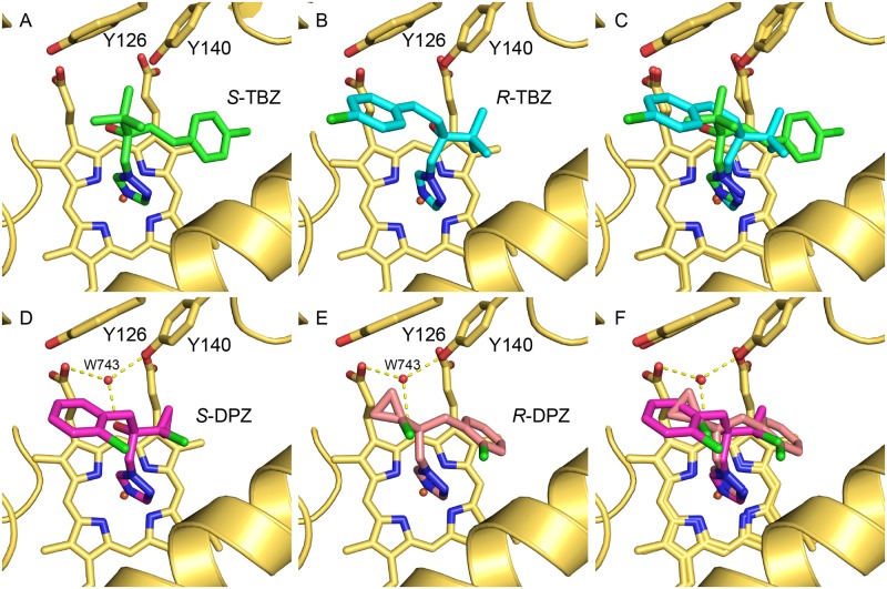
Fig 3. Phytopathogen inhibitors complexed in the active site of ScERG11p6×His.
The structures reveal the active site binding orientation of the enantiomers (A) S-TBZ (green carbons, PDB ID:5EAB), (B) R-TBZ (cyan carbons, PDB ID:5EAC), and (C) the superimposition of S-TBZ and R-TBZ, the active site orientation of the enantiomers (D) S-DPZ (magenta carbons, PDB ID:5EAD), (E) R-DPZ (salmon carbons, PDB ID:5EAE), and (F) the superimposition of S-DPZ and R-DPZ. The heme cofactor and selected residues (Y126 and Y140) are shown as sticks. Water-mediated (w743, red sphere) hydrogen bonds are shown as yellow dashed lines. Helix I is shown as a yellow ribbon at the bottom right of each panel. Nitrogen atoms are blue, oxygen red and chlorine green.

