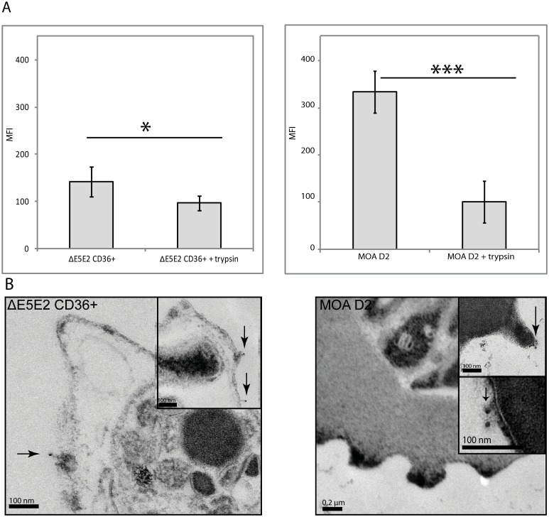
Fig 8. The MOA D2 surface recognition signal is trypsin sensitive.
(A) Surface recognition signal with day 70 sera before and after trypsinisation of CD36-selected ΔE5E2 (left panel) and of MOA D2 (right panel). Both ΔE5E2 and MOA D2 were incubated with MOA serum of day 70 and labelled with a secondary FITC antibody for detection in flow cytometry. Trypsinisation resulted in a significant decrease of the antibody recognition signal (standard errors are given, p = 0.05 for ΔE5E2 (n = 4) and p = 0.003 (n = 3) for D2) in both cell lines. (B) Immuno-TEM after MOA day 70 serum labelling on CD36 selected ΔE5E2 parasites and MOA D2 parasites. A secondary, 12 nm gold-labelled anti-human IgG antibody was added. In ΔE5E2 and MOA D2, gold particles (marked with arrows) could be detected on membrane areas with knobs as well as on the membrane areas without knobs.

