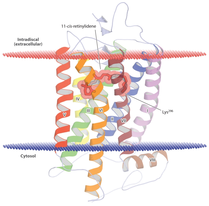
Figure 4.
Crystal structure of ground-state bovine rhodopsin. The protein backbone is shown in a schematic representation with the 11-cis-retinylidene chromophore and covalently linked lysine (Lys296) side chain displayed as sticks and spheres. The α-helices are numbered based on their order in the polypeptide sequence. The red and blue lines demarcate the approximate width of the phospholipid membrane bilayer. The figure was generated with PyMol (Schrödinger, https://www.pymol.org/) using the rhodopsin atomic coordinates deposited in the Protein Data Bank under accession code 1U19.

