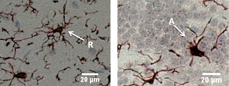
Figure 1.

Microglial morphology of rat olfactory bulb stained with anti-Iba1 antibody. Left, resting/ramified (R) microglia exhibiting thin, highly branched protrusions extending from the cell body; right, activated microglia (A) exhibiting amoeboid morphology with shorter, stouter processes. Left, rat treated with 110-nm silver nanoparticles 21 days post-exposure; right, rat treated with 20-nm silver nanoparticles 21 days post-exposure. Bar = 20 μm.
