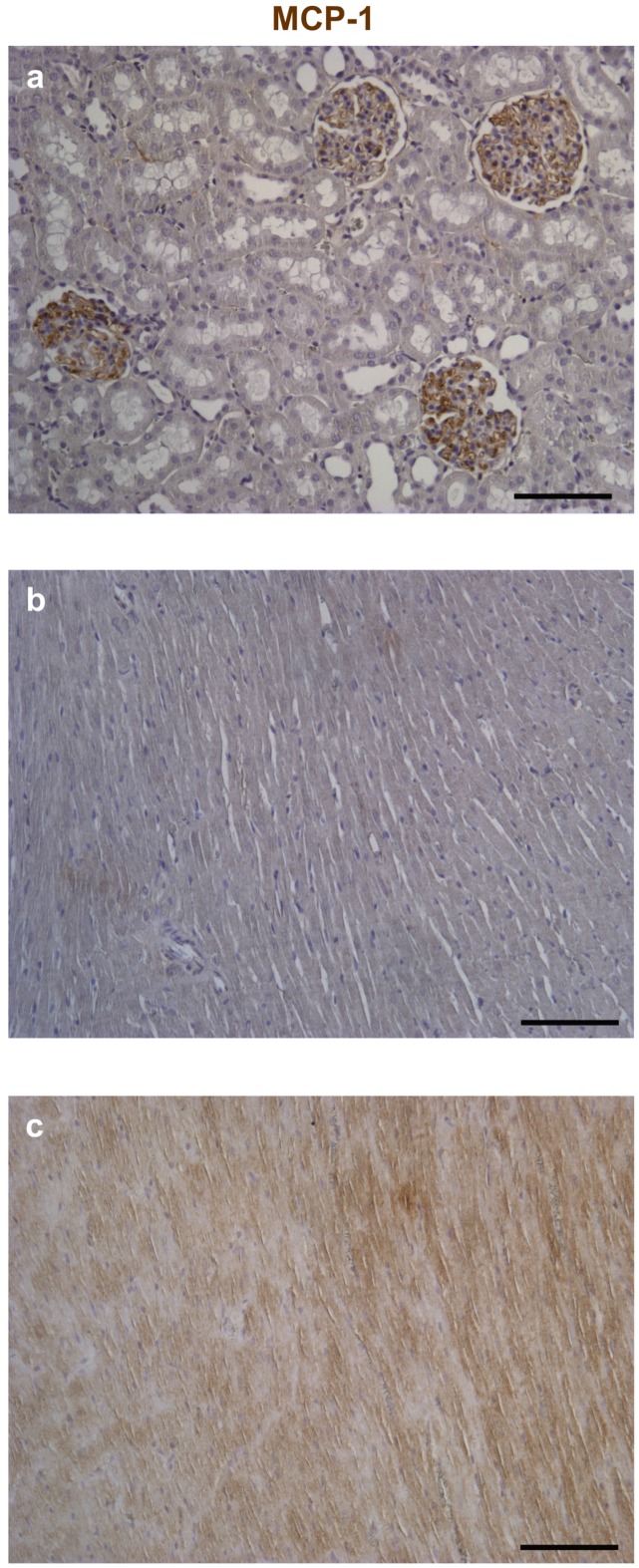
Figure 4.
Immunohistochemical analysis of Monocyte Chemoattractant Protein-1 (MCP-1). Reference tissues for the detection of MCP-1 by immunoperoxidase (brownish) were represented by the rat kidney (a); Compared to control (b); Higher expression of the chemokine is apparent in both cardiomyocytes and interstitial cells of the diabetic left ventricular (LV) myocardium (c). Nuclei were counterstained by Hematoxylin. Scale bars: 100 µm.

