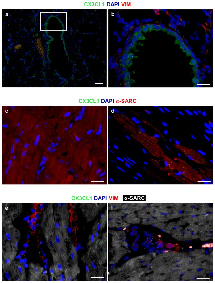
Figure 5.
Immunofluorescence analysis of CX3CL1 (Fractalkine). The granular and diffuse cytoplasmic expression of CX3CL1 (green) is apparent in bronchiolar epithelial cells of the rat lung, used as a positive control tissue; (a,b) The white square includes an area shown at higher magnification in b where interstitial cells are depicted by the red fluorescence of vimentin (VIM); (c) Internal negative control. Absence of immunofluorescence signals in a section of the rat diabetic heart exposed to FITC secondary antibodies in the absence of primary anti-CX3CL1 antibodies. Cardiomyocytes are recognized by the red fluorescence of alpha-sarcomeric actin (α-SARC); (d) A rather dense granular, dot-like pattern of CX3CL1 (green) expression in α-SARC (red) positive cardiomyocytes is illustrated in a section of the rat infarcted myocardium; (e,f) A granular, dot-like expression of Fractalkine (green) is apparent in VIM (red) positive fibroblasts and interstitial cells surrounding α-SARC (withe) positive cardiomyocytes. The pro-inflammatory cytokine is also detectable in cardiomyocytes. (a–f): Blue fluorescence corresponds to DAPI staining of nuclei. Scale Bars: (a) = 50 µm, (b–f) = 20 µm.

