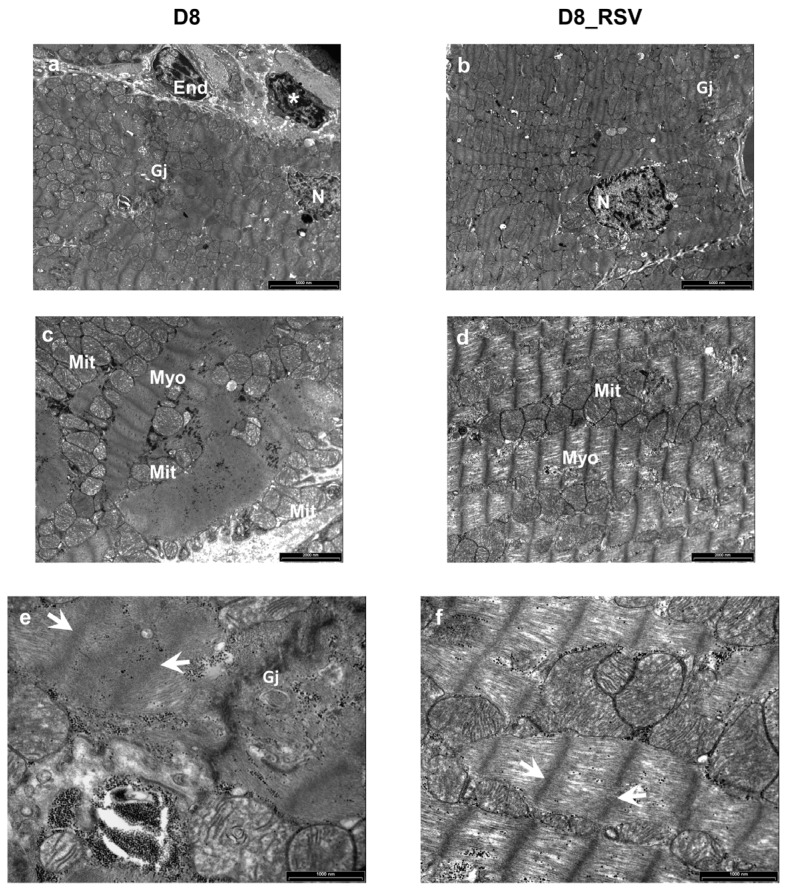
Figure 9.
Positive effects of RSV treatment on ultrastructural damage of diabetic LV myocardium. Ultra-thin longitudinal sections of the LV myocardium from untreated (D8) and RSV-treated (D8_RSV) diabetic rats. (a,b) Low magnification images illustrating cardiomyocytes connected by gap junctions (Gj). An inflammatory cell (*) showing electron-dense granules near a capillary endothelial cell (End) is also documented in the diabetic rat myocardium (a). N: cardiomyocyte nuclei. (c,d) Disorganization and severe alteration of contractile myofibrils (Myo) are illustrated in the diabetic myocardium (c) whereas a more preserved sarcomere striation is present in RSV-treated heart (d). Mitochondria (Mit) are running parallel to myofibrils in RSV-treated rat myocardium (d) which are swollen and clustering between disarrayed contractile filaments in diabetic rats (c). At higher magnification, a marked ultrastructural effacement of sarcomeres (white arrows) at the junctional interface (Gj) of diabetic cardiomyocytes (e) compared to RSV-treated myocardium (f) is apparent. Scale bars: (a,b): 5 µm; (c,d): 2 µm; (e,f): 1 µm.

