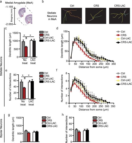
Figure 3. LAC oral prophylactic treatment rapidly promotes structural plasticity of MeA stellate, but not bipolar, neurons.
(a) Schematic of brain regional details along the dorso-ventral axis of the mouse brain with the bregma coordinates for the medial amygdala (MeA: highlighted in red), available from 2015 Allen Institute for Brain Science. Allen Mouse Brain Atlas26. (b) Representative tracing of Golgi-impregnated stellate neurons in the MeA. (c,e) CRS induced retraction and decreased the number of intersections of MeA stellate neurons, whereas 3 days of oral treatment with LAC, administered prior to the end of CRS, promoted structural plasticity of MeA stellate neurons (Length: F1,24=6.7, p<0.05 (treatment); F1,24=4.26, p<0.05 (stress); Intersections: F1,24=4.58, p<0.05 (treatment); F1,24=3.03, p=0.09 (stress)). (d,f) Morphometric Sholl analysis in incremental steps of 15μm from the soma shows that MeA stellate neurons underwent the most pronounced decrease in length and number of intersections in CRS mice starting from a distance of 105μm from the soma (see also SIFig.3). To note, Sholl analysis reveals that 3 days of LAC oral treatment promotes dendritic plasticity of MeA stellate neurons at the closer distance (75μm) from the soma (see also SIFig.3), suggesting that LAC prophylactic treatment induced growth of novel primary and secondary dendrites rather than reversing a pathological phenotype by elongation of CRS-shrink dendrites. (g,h) MeA bipolar neurons were not affected by CRS and LAC treatment as the total dendritic length and number of intersections appeared similar to non-stressed control mice. Bars represent mean+s.e.m.,*indicate significant comparisons with all of the other groups, *P<0.01.**P<0.001.

