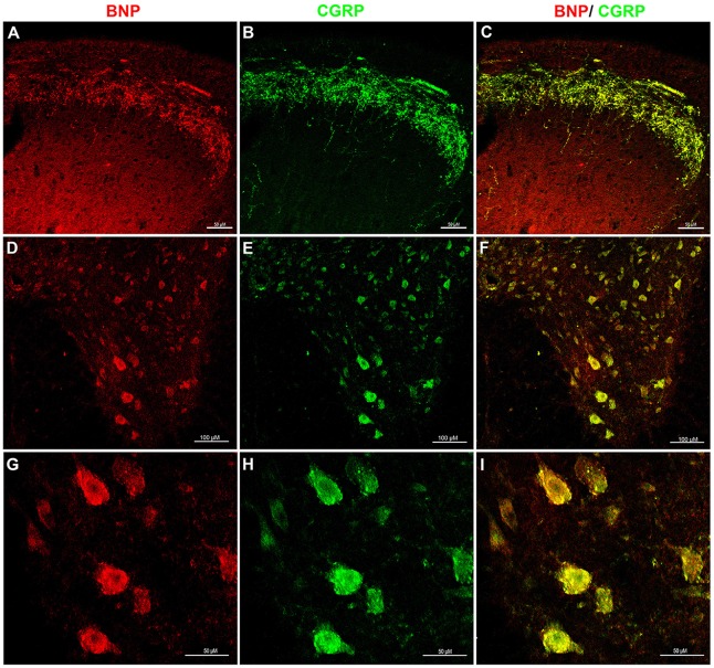
Figure 3.
Immunofluorescence confocal photomicrographs, showing the co-localization of BNP and calcitonin gene-related peptide (CGRP) in rat spinal cord. Spinal cord sections were labeled for BNP (red) and CGRP (green). (A) BNP immunoreactivity in the DH of the spinal cord. (B) CGRP-immunoreactive fibers in laminae I–II. (C) Merged images show the co-localization of BNP and CGRP in superficial laminae I–II. (D) A low-magnification image shows BNP immunoreactivity in the VH. (E) A low-magnification image shows CGRP immunoreactivity in the VH. (F) Merged images the co-localization of BNP and CGRP in the VH. (G) A high-magnification image shows BNP-immunoreactive neurons in the VH. (H) A high-magnification image shows CGRP-immunoreactive motor neurons in the VH. (I) Merged images show the co-localization of BNP and CGRP in the VH motor neurons. Scale bars in (A–C) and (G–I) = 100 μm and in (D–F) = 50 μm.

