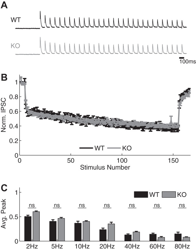
Fig. 8.

Hippocampal SC-associated inhibitory synapses exhibit unaltered STP in Fmr1 KO mice. A and B: sample traces (A) and normalized amplitudes (B) of IPSC recordings during 40-Hz stimulus trains in WT (black) and Fmr1 KO CA1 cells (gray) in the presence of DNQX (10 μM). Traces were normalized to the peak of the first response for visual comparison. C: normalized IPSC amplitudes at various frequencies in WT and Fmr1 KO neurons (ns, no significant difference).
